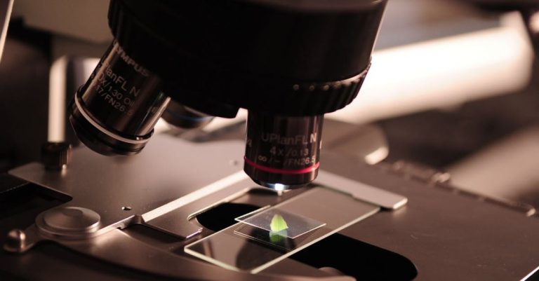
Visualizing the inner workings of living cells has been a fundamental goal in the field of biology for decades. With advancements in technology, live-cell imaging has revolutionized the way researchers study cellular processes in real-time. In recent years, there have been significant developments in live-cell imaging techniques that have opened up new possibilities for studying dynamic cellular events at high resolution. Let’s delve into some of the latest innovations in live-cell imaging that are shaping the future of biological research.
Super-resolution Microscopy: Breaking the Resolution Barrier
One of the most significant developments in live-cell imaging is the advent of super-resolution microscopy techniques. Traditional light microscopy has limitations in resolving structures smaller than the diffraction limit of light, approximately 200 nanometers. Super-resolution microscopy methods such as structured illumination microscopy (SIM), stimulated emission depletion microscopy (STED), and single-molecule localization microscopy (SMLM) have overcome this barrier, allowing researchers to visualize cellular structures at the nanoscale level. These techniques provide unprecedented detail and clarity, enabling scientists to study complex cellular processes with unparalleled precision.
Fluorescent Protein Technologies: Brightening Up Live-cell Imaging
Fluorescent proteins have become indispensable tools in live-cell imaging due to their ability to label specific cellular components and track dynamic events in real-time. Recent advancements in fluorescent protein technologies have led to the development of brighter, more photostable probes that enable longer imaging durations without photobleaching. Novel fluorophores with improved characteristics, such as increased brightness and faster maturation, have enhanced the quality of live-cell imaging data, allowing researchers to capture dynamic processes with higher sensitivity and resolution.
Live-cell CRISPR Imaging: Visualizing Gene Editing in Action
The revolutionary CRISPR-Cas9 gene editing technology has not only revolutionized the field of genetics but also opened up new possibilities for live-cell imaging. Live-cell CRISPR imaging involves fusing fluorescent proteins to the Cas9 enzyme, allowing researchers to track the editing process in real-time within living cells. This technique provides insights into the dynamics of gene editing events, such as DNA cleavage, repair, and target specificity, giving researchers a deeper understanding of the mechanisms underlying genome editing.
Microfluidics: Creating Controlled Environments for Live-cell Imaging
Microfluidic devices have emerged as powerful tools for live-cell imaging by providing precise control over the cellular microenvironment. These miniaturized systems enable researchers to manipulate the surrounding conditions of cells, such as nutrient availability, oxygen levels, and drug concentrations, while imaging cellular processes in real-time. Microfluidic platforms offer high spatial and temporal resolution, making them ideal for studying dynamic cellular events, such as cell migration, proliferation, and signaling, under controlled experimental conditions.
Machine Learning and Artificial Intelligence: Analyzing Big Data in Live-cell Imaging
As live-cell imaging generates vast amounts of data, the integration of machine learning and artificial intelligence (AI) has become essential for analyzing and interpreting complex image datasets. AI algorithms can automatically segment, track, and analyze cellular structures and behaviors in live-cell imaging videos, providing valuable insights into dynamic biological processes. By leveraging machine learning techniques, researchers can extract quantitative information from live-cell imaging data more efficiently and accurately, accelerating the pace of discovery in cell biology.
Innovations in live-cell imaging continue to push the boundaries of what is possible in biological research, allowing scientists to visualize and interrogate the intricate dynamics of living cells with unprecedented detail and clarity. With advancements in super-resolution microscopy, fluorescent protein technologies, live-cell CRISPR imaging, microfluidics, and artificial intelligence, the future of live-cell imaging holds immense promise for unlocking the mysteries of cellular life. As researchers continue to innovate and refine these techniques, the field of live-cell imaging is poised for exciting new discoveries that will reshape our understanding of the complexities of the cellular world.


